Under normal circumstances, the resumption of meiosis is triggered by LH surge which results in the degeneration of gap junctions through which meiosis resumption inhibitors are continuously delivered into the oocyte from corona cells. However, the resumption of meiosis can also be triggered mechanically removing corona cells which will eliminate the effects of meiosis inhibitors.
In the case of a postmature egg, it is apparent that a catastrophic event caused the degeneration of the corona cells and this was the most likely pathological trigger for the resumption of meiosis (if the egg was primed) or degeneration without the resumption of meiosis (if the egg was not primed). In the vast majority of cases, postmature eggs are sufficiently primed and will resume meiosis.
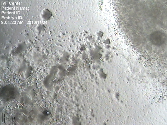
The cytoplasm of post-mature eggs, by its appearance and by stretching properties during ICSI, resembles the cytoplasm of unfertilized oocytes that were left in culture for about 2 days. Therefore, it is reasonable to speculate that a catastrophic event takes place about 2 days prior to hCG. Since this follicle was ready to resume meiosis about 2 days before hCG, it is further reasonable to speculate that this was one of the leading follicles.
In summary, a local (follicular) catastrophic event in one of the leading follicles that take place prior to hCG administration results in degeneration of cumulus cells, which leads to the resumption of meiosis in the oocyte that will have a post-mature appearance at the retrieval.
It is important to note that the affected follicle itself apparently persisted (since it could be aspirated), despite its content undergoing degenerative changes.
The link between premature luteinization and post-mature egg
The cumulus of post-mature oocytes exhibits signs of apoptosis rather than luteinization and usually, only a single egg in the cohort is affected. Therefore premature luteinization, which is caused by systemic factors can be ruled out, particularly because the premature elevation in LH is usually not observed in such patients. While the level of LH in these patients is normal, P4 may be elevated due to luteinization of some granulosa cells within the follicle with premature egg. This luteinization is spontaneous and apparently LH independent.
Possible link between premature luteinization and “vanishing follicles”
Vanishing follicles – follicles that spontaneously disappear during ovarian stimulation is a well-established phenomenon. It is particularly common in older patients, even though this may simply be due to the fact that in older patients it is easier to observe since the total number of follicles is small. In simple terms, those vanishing follicles simply “pop” when they reach a certain size, without any apparent help from any systemic hormones. This creates a possibility that they “pop” for simple mechanical reasons. Indeed, during the follicular phase, the volume of the follicle increases about 10 times. The surrounding tissues have to exhibit a considerable elasticity to accommodate such expansion. Furthermore, since the follicle is “local”, its expansion can be affected, for example, by ovarian scars from prior ovulations. Also, elasticity can vary from site to site and is probably lost with aging. Also, elasticity would be expected to vary from patient to patient.
Therefore, it is possible that some follicles originate from the area of the ovary where elasticity is lower and as its limit is reached, the follicle pops completely or, may lose its blood supply, in which case it would not pop, but persist, while its content, granulose cells die, triggering pathological oocyte maturation.
Since post-mature oocyte is apparently a local event, triggered at the level of the follicle, it is only affecting the potential of post-mature eggs, but no other eggs in the cohort.